For four years, a multidisciplinary team chased a map through a sliver of brain tissue smaller than a poppy seed, and on September 4 they released the finished version to the world: the complete wiring diagram of an adult male fruit fly, Drosophila melanogaster. Every neuron inside the brain is accounted for, along with each connection it makes, down to individual synaptic contacts.
The effort paired biologists at the Howard Hughes Medical Institute's Janelia Research Campus with computer scientists at Google, and the results appear in the journal Cell under the DOI 10.1016/j.cell.2026.08.015. It is the second complete Drosophila connectome, following a female fly brain map finished earlier in 2026 by a separate, largely university-based team. Together they give researchers matching reference brains of one species, male and female, drawn at synaptic resolution.
Getting from an intact brain sample to a finished diagram took roughly four years, and it leaned on tools that barely existed when the project began. The result is not a stylized cartoon but an itemized inventory of the brain's parts and wiring, a resource that other laboratories will interrogate for decades.
Key Facts
The adult fly nervous system holds roughly 150,000 neurons in total, and the new map covers the portion housed in the head, cataloging more than 300 million synaptic connections. A human brain, by contrast, contains about 86 billion neurons, more than half a million times as many. That gulf in scale is why Drosophila has become the proving ground for whole-brain connectomics: small enough to image exhaustively, yet complex enough to court, fight, learn and navigate.
To build the map, the team sliced the brain into ultrathin sections and imaged them for about a year with seven scanning electron microscopes at roughly eight nanometers of resolution, producing hundreds of terabytes of imagery. Artificial intelligence models stitched the slices into a coherent volume, traced the path of each neuron, recognized synapses, and predicted whether connections were excitatory or inhibitory. Human proofreaders then corrected the automated reconstruction by hand, a division of labor that made the project tractable.
Cell reported on September 4 that the reconstruction identifies more than 11,000 distinct cell types and traces circuits that span the whole brain rather than stopping at regional borders. Ars Technica reported on September 4 that the male map is the second complete fruit fly connectome and that its arrival lets researchers line up the sexes side by side. About 95 percent of the cells in the two brains are shared between males and females; the rest are sex-specific or dimorphic, present in both sexes but wired to different partners. Male-only interneuron classes form networks tied to courtship and aggression, and those differences cluster in higher-order regions rather than in the sensory and motor circuits both sexes rely on.
The most intriguing finding is small. A neuron that appears in the female connectome was absent from one hemisphere of the male brain but present in the other, a subtle asymmetry hinting that sex differences in behavior are encoded in fine-grained wiring details, not only gross architecture. The HHMI Janelia news release said on September 4 that discoveries like this are exactly what a complete map makes possible, because researchers can now ask precise questions about individual cells.
Analysis
What this really means is that neuroscience now has two complete reference brains for the same species, male and female, and the ability to compare them at single-neuron resolution. That is a new kind of scientific instrument. A researcher who wants to understand how a fly decides to fight or flee can now open a map, find the relevant cells, trace their inputs and outputs across the whole brain, and form hypotheses about how the behavior emerges from the wiring itself.
The sex comparison is the headline, but it deserves a note of caution. Sex differences in the fly are real and behaviorally consequential, yet they are a minority of what the connectome contains. Roughly 95 percent of the wiring is shared, so the two maps mostly confirm a common blueprint while highlighting the seams where the sexes diverge. Interpreting those seams is subtle: a cell that differs between males and females may be a cause of a behavioral difference, a consequence of one, or a developmental accident with no functional importance, and a static map cannot easily distinguish those possibilities.
The bigger picture here is that a connectome is a freeze frame. It captures the hardware of the brain at one moment in time, but not the software that runs on it. The map does not show which synapses are strong or weak, which neurons fire together, or how neuromodulators and experience reshape the circuits. The Google Research blog said on September 4 that the reconstruction is best understood as a foundation, one that becomes far more powerful when combined with recordings of neural activity, genetic tools that switch cells on and off, and behavioral experiments that test what the wiring predicts.
Even with those limits, a complete map compounds in value. Every future experiment on the fly brain, whether it involves optogenetics, calcium imaging or behavioral assays, can now be read against a known ground truth of connectivity. Circuits that once took years to reconstruct by hand can be queried across the whole brain in an afternoon. That changes the economics of neuroscience, and it is why the fly project was always about more than the fly.
Why It Matters
Drosophila earned its place in the laboratory because it breeds quickly, costs little to maintain, and comes with a century of genetic tools that let scientists delete or label individual genes with precision. Roughly 60 percent of human genes have recognizable counterparts in the fly genome, including a large share implicated in neurological disease. A wiring map of the fly brain is therefore not an entomological curiosity; it helps researchers interpret what those conserved genes do inside a working nervous system.
Timing matters as much as content. Connectomes have arrived in stages: a partial reconstruction known as the hemibrain, covering roughly 25,000 neurons, appeared in 2020, and the female whole-brain map followed in this cycle. Each step has grown larger, and the male map extends the project across the whole central nervous system while preserving the connections between regions that earlier efforts reconstructed separately.
The payoff is already visible in companion work. Teams have begun tracing complete pathways from sensory organs to the motor neurons that drive behavior, including the circuitry that turns taste into feeding decisions and the visual pathways a male fly uses to track a mate. Because the underlying dataset is open, any laboratory anywhere can browse the same volume of tissue and test its own hypotheses against the same ground truth, a departure from the days when such maps were closely held.
For human health, the relevance is indirect but real. No one will image a human brain at synaptic resolution soon, and the ethical and practical barriers are immense. But the fly project is where the methods are being invented and stress-tested. The imaging pipelines, machine learning models, proofreading strategies and data formats developed for Drosophila are the same tools that will eventually be aimed at larger brains, and every refinement lowers the barrier for the next species.
Next Up
The immediate next step is vertebrate. The network that built the fly map is already adapting its pipeline to larval zebrafish, whose small, transparent brain is far more tractable than a mammal's. Each jump in scale, from roughly 150,000 neurons in a fly to millions in a zebrafish larva and billions in a mouse, will demand faster imaging, more storage and better automated proofreading, but the trajectory is now clearly established.
In the nearer term, the two fly connectomes will keep yielding comparisons. Questions that were previously unanswerable become routine: which circuits are truly sex-specific, where do shared circuits diverge into different behaviors, and how much variation exists between individuals of the same sex. The asymmetry finding suggests that the answers will be found in fine detail rather than in broad strokes.
Companion studies published alongside the main map show where the field is heading. One lays out the complete taste pathway, following signals from sensory cells on the legs, wings and mouthparts to the motor circuits that drive feeding. Others examine how visual information reaches deeper into the brain than earlier models assumed, and how dimorphic neurons steer shared sensory input into sex-specific behavior. Together they illustrate the central promise of a connectome: a single reference that anchors an entire research program.
What comes after that is harder to predict, and that is rather the point. Past connectome releases, from worm to fly, tended to produce their most important discoveries years after publication, once the community had time to explore. The male fly brain map was built to be explored, and neuroscience is only beginning to learn its way around it.
Related Articles

Tsinghua one gram wireless microscope records over 8,500 neurons in moving mice
A Tsinghua led team has published details of a 1.0 gram head mounted mesoscope that images a 6 mm cortical field and captures more than 8,500 neurons in a freely moving mouse.
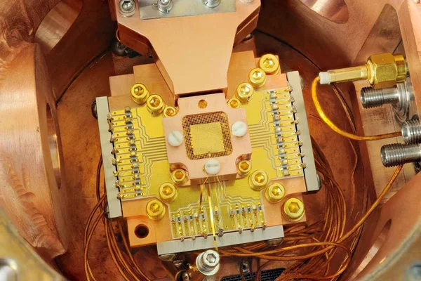
Quantinuum Hits 99.9995 Percent Logical Fidelity on Helios, a Tenfold Error Correction Record
The trapped-ion company says its 98-qubit Helios machine cut logical errors to five per million operations, a result outside researchers describe as a serious advance toward fault tolerance.
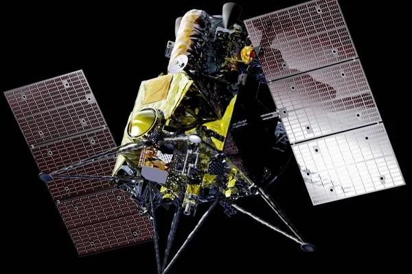
Japan set to launch MMX to Phobos on October 20 in world first Martian moon sample return
JAXA's Martian Moons eXploration spacecraft is scheduled to lift off from Tanegashima aboard an H3 rocket and aims to bring at least 10 grams of Phobos soil back to Earth by 2031.
Comments (0)
Log in or sign up to leave a comment.
No comments yet. Be the first to share your thoughts.